Les prix CIAPE soulignent la contribution à l’avancement des connaissances et l’investissement de nos membres dans la recherche sur les perturbateurs endocriniens, les méthodologies, les politiques ou la sensibilisation. Le 5 décembre 2024, quatre prix CIAPE ont été décernés au sein de notre communauté scientifique.
Cette conférence vous permettra d’en savoir plus sur les travaux de recherche de ces 4 membres du CIAPE.
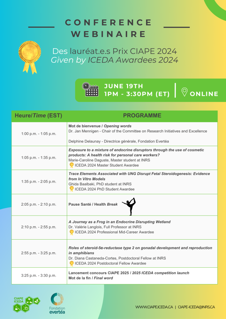
Détails
Date: 19 juin 2025, 13h – 15h30 (Heure de l’Est)
Lieu : Lien Zoom
Programme détaillé
Jen Mennigen – Présidente du Comité sur les initiatives et excellence en recherche.
Delphine Delaunay – Directrice Générale, fondation Evertéa
Marie-Caroline Daguste, Étudiante à la Maitrise, Institut national de la recherche scientifique
Résumé
Personal care workers (hairdressers, beauticians, manicurists) use many cosmetic products as part of their duties. Several endocrine disruptors (EDCs) have been identified in these products and studies have linked exposure to these compounds to various health problems. Our study aims to evaluate the effects of exposure to a mixture of EDCs in the workplace on breast health. To do this, 569 workers in personal care or in other fields (control group) responded to a questionnaire whose purpose was to establish a socio-economic profile of the participants as well as a profile of their exposure to EDCs. Preliminary analyses of the questionnaire indicated that the use of cosmetic products varied according to the participants’ occupation. This is significantly higher for the hairdresser group compared to the beautician group and the control group. Among the participants, 32 volunteers provided urine samples. Chemical analyses of these showed higher levels of parabens, benzophenones and bisphenols in the personal care workers group than in our control group. Finally, cytotoxicity and cell proliferation tests were also performed on tumorigenic (T47D) and non-tumorigenic (MCF-12A) mammary gland luminal cells that were exposed to urine samples. No significant effects on viability and mortality were observed for the 2 cell lines. The proliferation of T47D and MCF-12A cells does not appear to be affected by exposure to urine samples. This study will provide a better understanding of the risks associated with occupational exposure to EDCs.
Ghida Baalbaki, Étudiante au doctorat, Institut national de la recherche scientifique
Résumé
Biomonitoring studies have shown that pregnant women living in regions of unconventional natural gas (UNG) exploitation have higher levels of trace elements. Whether developmental endocrine disruption can be expected at these exposure levels during pregnancy is unclear. In this study, we aimed to test the impact of five trace elements alone or in mixtures using in vitro cell- and tissue-based assays relevant to endocrine disruption and development. Manganese, aluminum, strontium, barium, and cobalt were tested at concentrations including those representatives of human fetal exposure. Using transactivation assays, none of the tested elements nor their mixture altered the human estrogen receptor 1 or androgen receptor genomic signalling. In the rat fetal testis assay, an organ culture system, cobalt (5 μg/l), barium (500 μg/l) and strontium (500 μg/l) significantly increased testosterone secretion. Cobalt and strontium were associated with hyperplasia and/or hypertrophy of fetal Leydig cells. Mixing the five elements at concentrations where none had an effect individually stimulated testosterone secretion by the rat fetal testis paralleled by the significant increase of 3β-hydroxysteroid dehydrogenase protein level in comparison to the vehicle control. The mechanisms involved may be specific to the fetal testis as no effect was observed in the steroidogenic H295R cells. Our data suggest that some trace elements in mixture at concentrations representative of human fetal exposure can impact testis development and function. To further assess effects on feto-placental steroidogenic communication, we will use a human co-culture model combining BeWo (trophoblast-like) and H295R (fetal-like) cells, which mimics hormone production during pregnancy. This model will allow us to investigate the endocrine disrupting potential of trace elements on key pregnancy hormones and placental aromatase activity. This study highlights the potential risk posed by UNG operations, especially for the most vulnerable populations, pregnant individuals, and their fetus
Dr. Valérie Langlois, Professeur Titulaire, Institut national de la recherche scientifique
Résumé
This presentation will feature Dr. Langlois’ major contributions to the field of endocrine disruption over the last decades. It will emphasize key initiatives she has led and provide an overview of her extensive research program. Her work focuses on the effects of various endocrine disrupting chemicals (EDCs) on amphibians, particularly frogs. These chemicals include phthalates, bisphenol A, arsenic, atrazine, disperse yellow 7, diluted bitumen, finasteride, and ifosfamide, among others. Dr. Langlois’ primary research areas involve the disruption of multiple hormonal axes, including androgen, thyroid, stress, progesterone, and prolactin systems. The second part of the talk will delve into her current research on the impact of melengestrol acetate, highlighting novel regulatory mechanisms observed in frogs and their broader implications for environmental and endocrine research. The presentation will showcase the work of her trainees and collaborative efforts within her team.
Dr. Diana Castaneda-cortes, Stagiaire postdoctorale, Institut national de la recherche scientifique
Résumé
The enzyme steroid-5α-reductase type 2 (Srd5α2) is crucial in the biosynthesis of 5α-dihydrotestosterone (5α-DHT), one of the most potent androgens in vertebrates. Given the increasing prevalence of environmental contaminants that act as endocrine disruptors in both animal and human populations, it is essential to investigate the regulation and production of androgens to elucidate the health implications associated with altered androgen biosynthesis. In this study, we utilized CRISPR gene-editing technology to create frog mutants (srd5α2+1△4) of the Western clawed frog (Xenopus
tropicalis) to investigate the consequences of androgen disruption. Our research focuses on characterizing the roles of Srd5α2 in gonadal development and differentiation, as well as its influence on reproductive capacity and secondary sex characteristics. Our findings reveal that over 80% of the homozygous male mutants exhibited significant reductions in nuptial pad development, an androgen-dependent secondary sexual characteristic, compared to wild-type males. Additionally, histological analysis of gonadal maturation shows a deficient spermatogenesis in mutants. This is demonstrated by the presence of empty cysts during climax metamorphosis and one month post-metamorphosis. As a result, by the time the individuals reach sexual maturity (three months post-metamorphosis), over 40% of those mutants exhibited intersex gonads. We also examined the molecular compensation mechanisms related to the other types of 5-reductases (srd5α1, srd5α3, and srd5β) and the androgen receptor (ar) during embryonic sex determination. It was found that both srd5β and ar were up-regulated in the mutants, whereas the expression levels of srd5α1 and srd5α3 were like those observed in wild-type embryos. While the precise role of 5β-dihydrotestosterone in gonadal development and reproduction is not fully understood, this study provides important insights and highlights the need for additional research into the regulation of androgen production in aquatic and semi-terrestrial species. Acknowledgments: Marko Horb (National Xenopus Resource (NXR), Marine Biological Laboratory).